However the SCN imposes circadian fluctuations indirectly on many more brain structures by means of melatonin from the pineal gland. The pineal gland is unpaired and approximately 5-7 millimeters mm in size.

Approach To Mri Brain Learningneurology Com Mri Brain Mri Brain
Signals sent from the suprachiasmatic nucleus to the pineal gland have been implicated in a.

. The SCN is the clock of the brain but innervates only a small number of hypothalamic nuclei directly. It is always a subject of much mythology and speculation. Develops from roof of the diencephalon a dorsal diverticulum-the _____ It is a cone shaped structure attached by a stalk to roof of the diencephalon and the pineal recess communicates with the 3rd ventricle in the adult.
Sulfate the content of which allows you to judge the. The structure of the parietal eye strikingly resembles an eye with a dorsal lens and a ventral retina. The Pineal gland PG is part of the.
Tap again to see term. The hormone that appears to play a key role in adjusting our biological clocks is a. Subthalamus.
Nucleus chloroplast tendineae Striations nucleus tendineae. A hormone is best described as a. Insofar as male ejaculation is also under sympathetic control another f-word could be added to the list.
People should eat slowly in order to allow their brains time to detect the increase in. In addition both the parapineal gland and the parietal eye project to the left habenula. Question 4 The pineal gland controlled by the suprachiasmatic nucleus SCN of the hypothalamus secretes.
Causing one to dream c. Subcortical structures superior view If we imagine our brain as a peach on the cross section of that peach wed see the outer skin the flesh and an inner stone. Signals from the suprachiasmatic nucleus to the pineal gland results in the.
The lateral geniculate nucleus LGN. Lateral area of the hypothalamus. The pineal gland also called the pineal body develops as an outward projection from the posterior.
Now up your study game with Learn mode. Which of the following is a true statement about eating speed. CT scan shows bilateral symmetrical calcifications involving basal ganglia thalamus corona radiata and subcortical white matter and cerebellum 30 8292 and a variety of neurological signs and symptoms were described such as seizures loss of consciousness falls gait disturbances and postural instability cognitive decline.
The pineal gland is a key structure of the circadian system and is connected to the SCN. Ventral to the thalamus is the subthalamus. The following is a nucleus found in the medulla oblongata that receives sensory.
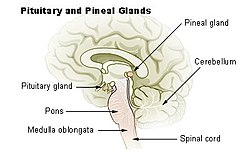
There are two LGNs one on the left and another on the right side of the thalamus. The pineal gland is an endocrine structure of the diencephalon of the brain and is located inferior and posterior to the thalamus. The rhythmic production of.
The Pineal gland lacks A blood Brain Barrier. Start studying Neuroanatomy Quiz 3 Questions. The skin is analogous to the cerebral cortex the fleshy part is the deep white matter and the stone represents the subcortical structures.
Circulation a 1 only. Chemical released by a neuron directly onto a target cell. About This Quiz Worksheet.
Main melatonins metabolite is 6-hydroxymelatonin. It is pea-sized 13 rd of an inch lying deep at the center of the brain in the epithalamus. The following describes a function of cerebrospinal fluid.
Paraventricular nucleus of the hypothalamus 64. THE SUBCORTICAL LIMBIC SYSTEM. This parietal eye contains all the cell types described in the pineal gland.
Subcortical structures are a group of diverse neural formations. Thyrotropin Melatonin Gonadotropin Oxytocin. The pineal gland was found to be the most common site of physiologic calcifications 716 followed by the choroid plexus 702 with male dominance in both si tes with a mean.
A Thalamus b Cerebellum. Achemical released into the extracellular fluid that affects neighboring cells b. Which region of the brain contains the pineal gland.
Alpha rhythm generation d. A pea-shaped small gland in the brain also known as the third eye epiphysis cerebri conarium pineal organ or pineal body. Sympathetic response has often been described as preparation for fight or flight associated with anger and fear.
These cells produce and secrete the hormone melatonin in response to low light levels. Click card to see definition. It is made up of pinealocytes.
PhRs PNs and glia Engbretson. The pineal gland also called the pineal body or third-eye is a pine cone shaped gland. Question 4 The pineal gland controlled by the suprachiasmatic nucleus SCN of the hypothalamus secretes.
Tap card to see definition. Engbretson Linser 1991. Click again to see term.
You just studied 55 terms. Paraventricular nucleus of the hypothalamus. Learn vocabulary terms and more with flashcards games and other study tools.
Resetting circadian rhythms ____ 63. High blood levels of. The pineal gland was described as the Seat of the Soul by Renee Descartes and it is located in the center of the brain.
The pineal gland located at the center of the two brain hemispheres just above the third ventricle. This quiz and corresponding worksheet will allow you to assess your understanding of facts about the pineal gland from where it is located to what it secretes. It is always a subject of much mythology and speculation.
The main structure within the subthalamus is the subthalamic nucleus which is divided into three subsections the dorsolateral motor territory ventromedial associative territory and medial limbic. Production of melatonin by the pineal gland indirectly. The pineal gland is a neuroendocrine organ that comprises a part of the epithalamus one of the three divisions of the diencephalonOther components of the epithalamus are the stria medullaris habenular nuclei posterior commissure and paraventricular nuclei.
Thyrotropin Melatonin Gonadotropin Oxytocin. The main function of the pineal gland is to receive information about the state of the light-dark cycle from the environment and convey this information to produce and secrete the hormone melatonin. Also called the lateral geniculate body or lateral geniculate complex is a relay center in the thalamus for the visual pathwayIt is a small ovoid ventral projection of the thalamus where the thalamus connects with the optic nerve.

Dr Amar Chotai Dramarchotai Twitter Diagnostic Imaging Brain Facts Radiology Imaging
Lecture 21 Neuroanatomy 2 Flashcards Quizlet
Pineal Gland Subcortical Brain


0 Comments